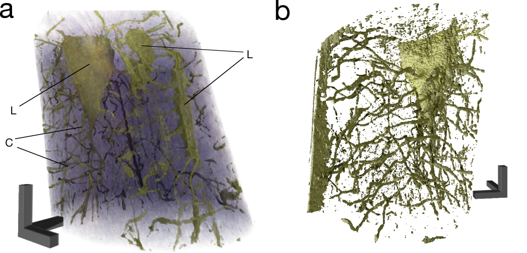
High-resolution three-dimensional digital image of the bone sample
Image explanation:
The specially developed nano-CT algorithm uses the multiple X-ray images (indicated on the left) to compute a high-resolution three-dimensional digital image of the sample. Applied in osteology, the process helps to visualize the fine network of channels about 100 nanometers in size, through which cell extensions connect the various bone cells with each other.